AI for radiograph analysis integrates at three key points in your existing workflow: image capture, review and diagnosis, and data population. After a sensor or phosphor plate captures an image, it is typically saved to your imaging software (e.g., Dexis, Schick) and linked to the patient's chart in your PMS (Dentrix, Eaglesoft, etc.). At this moment, an AI service can be triggered via a secure API call or a watched folder to analyze the DICOM or image file. The AI processes the radiograph for pathologies like caries, bone loss, calculus, and periapical lesions, returning structured findings—not just a PDF report, but discrete data points ready for your PMS.
Integration
AI Integration for Dental Radiograph Analysis AI
A technical guide to integrating AI-powered radiograph analysis directly into dental practice management systems, automating pathology detection and populating structured clinical findings.
FROM CAPTURE TO CHART
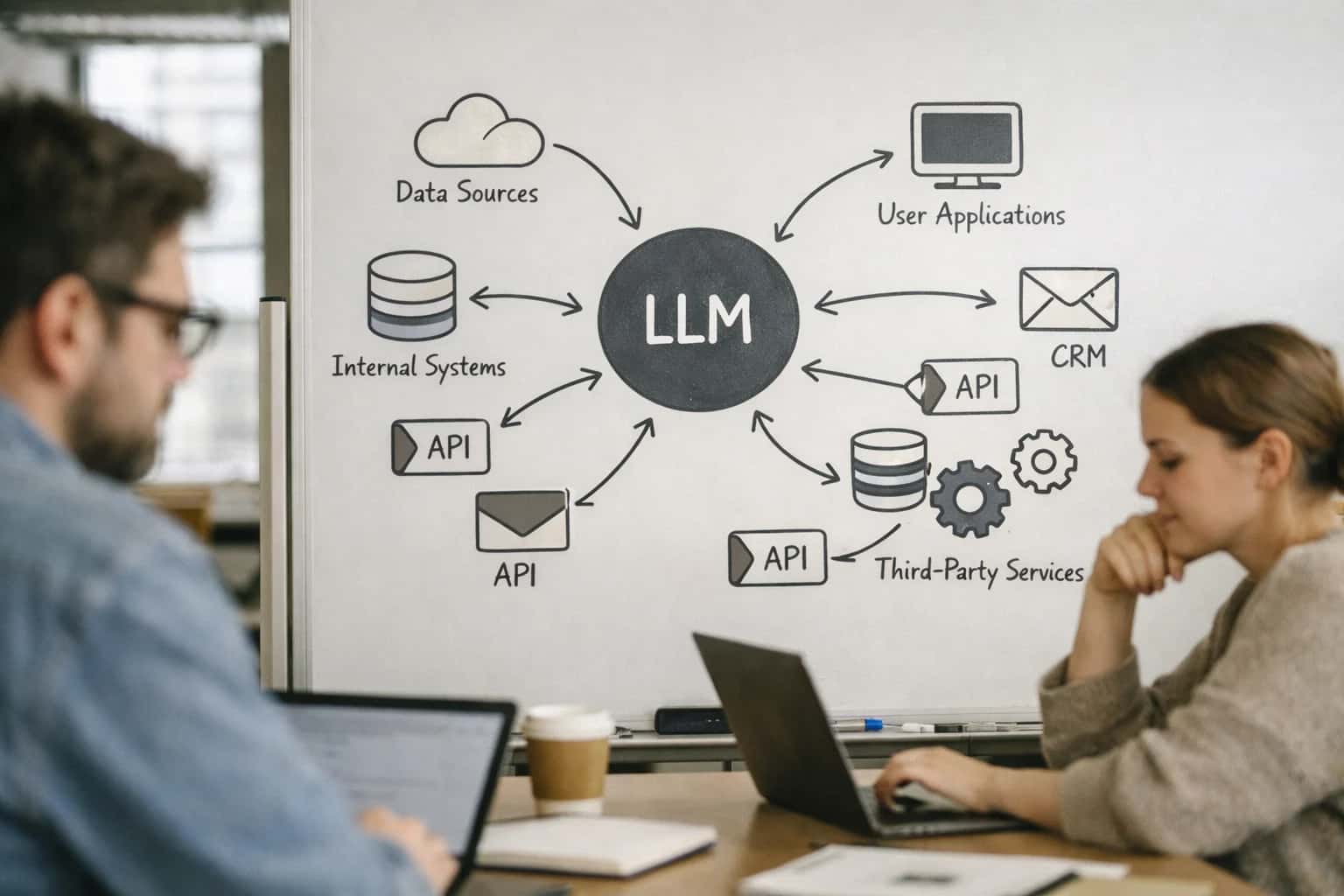
Where AI Fits in the Dental Radiograph Workflow
A practical blueprint for integrating AI analysis into the existing image management and charting workflow of your dental practice management system.
The high-value integration is the automatic population of structured data fields in the patient's clinical chart. Instead of a dentist manually typing observations, the AI's findings—such as 'MOD caries on tooth #19, distal surface' or 'horizontal bone loss >30% on #24-26'—can be written back to designated custom fields or note templates within the PMS charting module. This creates an auditable, searchable record. For governance, the system should log the AI inference, require a dentist's review and sign-off on the findings before final chart entry, and maintain the original radiograph as the source of truth. This workflow reduces charting time from minutes to seconds and ensures consistent documentation language.
Rollout is typically phased. Start with non-diagnostic support, like auto-tagging images with tooth numbers and view types to organize the image library. Then, pilot assistive diagnostics in hygiene exams for preliminary charting, where the hygienist and dentist review AI-highlighted areas. Finally, implement production diagnostics for specific, high-confidence use cases like caries detection on bitewings. The integration architecture is lightweight: a secure cloud service (or on-premise appliance) acts as a bridge between your imaging software's export function and your PMS's API, handling PHI compliance, result queuing, and audit trails. This approach augments your team's expertise without disrupting the familiar flow of opening an image, making a diagnosis, and updating the chart.
AI FOR RADIOGRAPH ANALYSIS
Integration Touchpoints in Dental PMS & Imaging Systems
Connecting AI to the Imaging Pipeline
The primary integration surface is the Picture Archiving and Communication System (PACS) or the imaging module within the PMS (e.g., Dexis, Schick, Sidexis). AI analysis is triggered at two key points:
- Post-Capture Ingestion: When a new radiograph (bitewing, periapical, panoramic, CBCT) is saved to the PACS, a webhook or API call sends the DICOM file to the AI inference service. This enables real-time, background analysis without disrupting the clinical workflow.
- Pre-Review Enrichment: Before the dentist opens the study, the AI service returns structured findings (e.g.,
potential_caries: MOD_surface_#19,bone_loss_severity: moderate,findings_confidence: 0.92). These results are stored as metadata or in a linked findings table, ready to populate the clinical note.
Integration ensures the AI acts as a silent assistant, annotating studies for review rather than replacing clinical judgment.
INTEGRATED WITH DENTAL PRACTICE MANAGEMENT
High-Value AI Radiograph Analysis Use Cases
Integrating AI-powered radiograph analysis directly into your dental PMS (Dentrix, Eaglesoft, Open Dental, Curve) transforms static images into structured, actionable data. These use cases show how findings automatically populate patient charts, trigger workflows, and enhance clinical decision-making.
01
Automated Caries Detection & Charting
AI analyzes bitewing and periapical X-rays to flag potential decay areas. Findings, including lesion location and suggested ICD-10/SNOMED codes, are written directly to the structured data fields in the PMS patient chart, pre-populating the treatment plan module for review.
Batch → Real-time
Analysis speed
02
Periodontal Bone Loss Quantification
For perio patients, AI measures bone levels on full-mouth series or bitewings, calculating bone loss percentages per tooth. Results automatically update the periodontal chart in the clinical module, providing a longitudinal view for monitoring disease progression and hygiene recall planning.
Same day
Chart update
03
Impacted Tooth & Pathology Triage
AI reviews panoramic and CBCT scans to identify impacted teeth, cysts, or other pathologies. A summary report with annotated images is attached to the patient's document management module in the PMS, and an alert can be created in the clinical notes to prompt specialist referral workflows.
Hours → Minutes
Review time
04
Pre-Treatment Implant Site Analysis
Prior to implant planning, AI assesses CBCT scans for bone density, nerve canal proximity, and sinus location. Key measurements and a risk assessment are appended to the patient's treatment plan in the PMS, providing the surgeon with integrated data for case presentation and consent.
1 sprint
Integration timeline
05
Orthodontic Cephalometric Tracing
For orthodontic practices, AI performs automated landmark identification and tracing on cephalometric X-rays. Calculated angles and measurements (SNA, SNB, etc.) are pushed into the patient's orthodontic module within the PMS, providing instant data for diagnosis and treatment planning.
Batch → Real-time
Tracing workflow
06
Longitudinal Change Detection
AI compares current radiographs with prior studies in the PMS image library to detect subtle changes in existing lesions, bone levels, or healing progress. A change report is generated and linked to the clinical note, supporting preventive care recommendations and insurance narrative justifications.
PRODUCTION IMPLEMENTATION PATTERNS
Example AI-Enhanced Radiograph Workflows
These workflows illustrate how AI analysis of dental radiographs integrates with your practice management system (PMS) to automate findings, populate structured data, and trigger clinical or administrative actions. Each pattern is designed to augment, not replace, the dentist's final review.
Trigger: A new bitewing radiograph series is saved to the patient's document tab in the PMS (e.g., via direct integration with Dexis, Schick, or a DICOM router).
Context Pulled: The PMS API is called to retrieve the patient's age, last prophylaxis date, and existing caries/restoration history.
AI Agent Action:
- The AI model analyzes the new radiographs for:
- Interproximal caries (scoring lesion depth: E0, E1, E2, D1, D2, D3)
- Existing restoration margins
- Alveolar bone levels
- A structured JSON finding is generated:
json{ "study_type": "bitewing", "findings": [ { "tooth": "3", "surface": "M", "finding": "recurrent_caries", "confidence": 0.92 }, { "tooth": "30", "surface": "D", "finding": "bone_loss", "severity": "mild", "confidence": 0.87 } ], "summary": "Potential recurrent caries on #3M; mild bone loss noted on #30D." }
System Update:
- The summary is appended as a draft note in the clinical notes module, tagged for hygienist/dentist review.
- Structured findings populate discrete fields in a custom "AI Findings" tab within the patient chart.
- If caries confidence exceeds a threshold (e.g., >0.9), a suggested treatment code (D0230, D2140) is pre-loaded in the treatment plan module.
Human Review Point: The hygienist and dentist review the AI findings during the exam, confirming or editing them before signing the note and finalizing the treatment plan.
FROM PACS TO PMS
Implementation Architecture: Data Flow & APIs
A secure, event-driven architecture to connect AI analysis engines with your dental practice management system, turning radiographic findings into actionable clinical data.
The integration is built on a secure API gateway that acts as a bridge between your imaging system (PACS) and your Practice Management Software (PMS). When a new radiograph is saved in your PACS (e.g., Dexis, Schick), a webhook event is sent to a dedicated ingestion service. This service fetches the anonymized DICOM image, passes it to the AI analysis model (hosted in a HIPAA-compliant cloud or on-premises), and receives a structured JSON report detailing findings like caries, bone loss, or periapical pathologies. The critical step is mapping these findings to the correct patient and visit in your PMS (Dentrix, Eaglesoft, Open Dental, or Curve) using a unique study ID or patient identifier passed in the initial event.
Once the AI analysis is complete, the integration uses the PMS's native REST or SOAP API to update the patient's clinical record. This typically involves: creating a new clinical note or progress note entry that summarizes the AI findings in plain language; attaching the original and annotated images to the patient's document module; and, most importantly, populating structured data fields. For example, surface-level caries detection can auto-populate tooth-specific charting surfaces, while bone level measurements can update periodontal charting data. This creates an auditable link between the AI's output and the official patient record without manual data entry.
Governance is managed through a central orchestration layer that handles error recovery, audit logging, and optional human-in-the-loop review. For high-confidence findings, updates can be applied automatically. For ambiguous cases, the findings can be routed to a review queue within the PMS's task module for dentist verification before chart updates are committed. All API calls are logged with user context (either system service or reviewing dentist) for full traceability. Rollout typically starts with a single operatory or specific procedure type (e.g., bitewing analysis) to validate data flow and clinical acceptance before scaling to full practice adoption.
RADIOLOGY WORKFLOW INTEGRATION
Code & Payload Examples
Automating Radiograph Intake
When a new radiograph is saved in your imaging software (e.g., Dexis, Schick), a webhook can trigger an AI analysis pipeline. The first step is to securely transfer the DICOM or image file, tag it with patient and visit metadata from the PMS, and queue it for processing. This payload example shows the event sent from the PMS to initiate the workflow.
json{ "event_type": "new_radiograph", "pms_patient_id": "DENTRIX-12345", "pms_visit_id": "APT-2024-05-15-09:00", "image_url": "https://secure-pacs.example.com/studies/1.2.840.113619.2.404.3.27985062.102.1715785200.123", "image_type": "bitewing", "laterality": "quadrant_3", "metadata": { "provider_id": "DR-SMITH", "date_of_service": "2024-05-15", "procedure_code": "D0274" } }
The AI service receives this payload, retrieves the image, and processes it through pathology detection models for caries, bone loss, calculus, and other findings.
RADIOGRAPH ANALYSIS INTEGRATION
Realistic Time Savings & Operational Impact
How integrating AI analysis for dental radiographs changes daily workflows and operational metrics within your practice management system.
| Metric | Before AI | After AI | Notes |
|---|---|---|---|
Radiograph Review & Initial Findings | 5-10 minutes per full-mouth series | 1-2 minutes for AI-generated summary | Dentist reviews AI-highlighted areas; findings auto-populate chart notes |
Pathology Documentation in Chart | Manual note entry from visual scan | Structured data auto-inserted into PMS fields | AI suggests standardized codes (e.g., for caries, bone loss) for review |
Treatment Plan Data Assembly | Gather images, notes, and history manually | AI pre-assembles relevant data into a draft plan | Clinical decision support provides evidence-based context for plan |
Patient Consultation Prep | 15-20 minutes to compile visuals and explanations | 5 minutes to review AI-generated patient report | Report includes annotated images and layperson explanations from PMS data |
Insurance Pre-authorization Support | Manual annotation of radiographs for justification | AI auto-generates clinical justification notes | Notes are formatted for common payer requirements and attached to claim |
Follow-up Scheduling & Recall Triggers | Manual review of charts to flag needed follow-ups | AI flags patients needing follow-up based on findings | Triggers automated recall workflow in PMS scheduling module |
Quality Assurance & Peer Review | Random manual audit of radiographic interpretations | AI provides consistency scoring across provider interpretations | Helps identify training opportunities; audit trail stored in PMS |
IMPLEMENTING AI IN A REGULATED CLINICAL ENVIRONMENT
Governance, Security & Phased Rollout
Deploying AI for radiograph analysis requires a secure, governed approach that integrates with existing clinical workflows and compliance frameworks.
A production integration connects the AI analysis service to your image management system (PACS) and Practice Management System (PMS) via secure APIs. The typical flow is event-driven: when a new radiograph is saved in the PACS, a secure webhook notifies the AI service. The service retrieves the anonymized DICOM image via a zero-trust API, processes it, and returns structured findings—such as potential caries, bone loss percentage, or periapical pathology—as a JSON payload. This payload is then written to pre-defined structured data fields in the patient's dental chart within the PMS (e.g., Dentrix Progress Notes, Eaglesoft Clinical Module), creating an auditable link between the original image and the AI-generated observation.
Governance is built around human-in-the-loop review and audit trails. Findings are presented to the dentist as draft annotations or notes, requiring a clinician's review and sign-off before being finalized in the patient record. All AI interactions are logged with a full audit trail: original image ID, model version used, inference timestamp, reviewing clinician, and final action taken. This satisfies clinical responsibility requirements and creates a dataset for continuous model validation and improvement. Access is controlled via the PMS's existing Role-Based Access Control (RBAC), ensuring only authorized clinical staff can view or act on AI suggestions.
A phased rollout minimizes disruption and builds trust. Phase 1 (Pilot): Integrate with a single operatory or hygienist column for bitewing analysis only. Findings are delivered to a separate review dashboard, not the live chart. Phase 2 (Expansion): Enable auto-population of structured fields in the PMS chart for approved findings, expanding to periapical and panoramic X-rays. Phase 3 (Scale): Roll out to all providers, introduce predictive alerts for high-risk findings, and connect insights to patient communication workflows for automated preventive care reminders. Each phase includes feedback loops where dentists can flag incorrect analyses, which are used to refine prompts and improve model performance specific to your practice's patient demographics and imaging equipment.
Enabling Efficiency, Speed & Accuracy
Intelligent Analysis, Decision & Execution
We build AI systems for teams that need search across company data, workflow automation across tools, or AI features inside products and internal software.
Talk to Us
Search across company data
Give teams answers from docs, tickets, runbooks, and product data with sources and permissions.
Useful when people spend too long searching or get different answers from different systems.
Enterprise searchRAGPermissions
Read more
Automate internal workflows
Use AI to route work, draft outputs, trigger actions, and keep approvals and logs in place.
Useful when repetitive work moves across multiple tools and teams.
AI agentsWorkflow automationGovernance
Read more
Add AI to products and internal tools
Build assistants, guided actions, or decision support into the software your team or customers already use.
Useful when AI needs to be part of the product, not a separate tool.
AI integrationDecision supportModel routing
Read moreIMPLEMENTATION BLUEPRINT
Frequently Asked Questions
Practical questions for integrating AI radiograph analysis into your dental practice management system, covering workflows, data handling, and rollout.
The integration acts as a secure bridge between your imaging system (PACS) and your Practice Management System (PMS).
Typical Data Flow:
- Trigger: A new radiographic study is saved in your imaging software (e.g., Dexis, Schick).
- Ingestion: A secure listener (webhook or scheduled poll) detects the new DICOM image and sends it to the AI analysis service.
- Analysis: The AI model processes the image, detecting pathologies like caries, bone loss, periapical lesions, and calculus.
- Structured Output: Findings are returned as structured JSON data, including pathology type, location, confidence score, and a textual summary.
- PMS Update: The integration service calls the PMS API (e.g., Dentrix, Eaglesoft) to:
- Attach the AI findings report as a document to the patient's chart.
- Populate structured data fields (e.g., custom clinical observations or perio charting fields) with quantified results.
- Create a follow-up task or alert for the dentist's review if high-confidence findings are present.
Key Architecture: This typically uses a cloud-based microservice that never stores PHI, acting as a stateless processor with audit logs for all image access.

About the author
Prasad Kumkar
CEO & MD, Inference Systems
Prasad Kumkar is the CEO & MD of Inference Systems and writes about AI systems architecture, LLM infrastructure, model serving, evaluation, and production deployment. Over 5+ years, he has worked across computer vision models, L5 autonomous vehicle systems, and LLM research, with a focus on taking complex AI ideas into real-world engineering systems.
His work and writing cover AI systems, large language models, AI agents, multimodal systems, autonomous systems, inference optimization, RAG, evaluation, and production AI engineering.
LinkedIn
Limited slotsGet a Free AI Consultation
Partnered with leading AI, data, and software stack.


How We Work
Custom AI workflows for your Business
One-fit-all AI don't work for modern businesses. At Inferensys, we aim to understand your business & custom requirements; which we use to define most efficient agentic workflows, the data, and the tools for your business.
01
Review the use case
We understand the task, the users, and where AI can actually help.
Read more02
Pick the right approach
We define what needs search, automation, or product integration.
Read more03
Build the first useful version
We implement the part that proves the value first.
Read more04
Improve from there
We add the checks and visibility needed to keep it useful.
Read moreThe first call is a practical review of your use case and the right next step.
Talk to Us