Cross-scanner harmonization is a computational technique that mitigates the non-biological variance introduced by different MRI or CT scanner manufacturers, magnetic field strengths, or reconstruction algorithms. By applying methods like CycleGAN adaptation or Hounsfield Unit normalization, it transforms images from disparate source domains to match a reference target domain, effectively reducing domain shift at the pixel or feature level.

Glossary
Cross-Scanner Harmonization
The computational process of normalizing medical images acquired from different scanner vendors or protocols to eliminate site-specific intensity variations, enabling robust, generalizable AI model training.
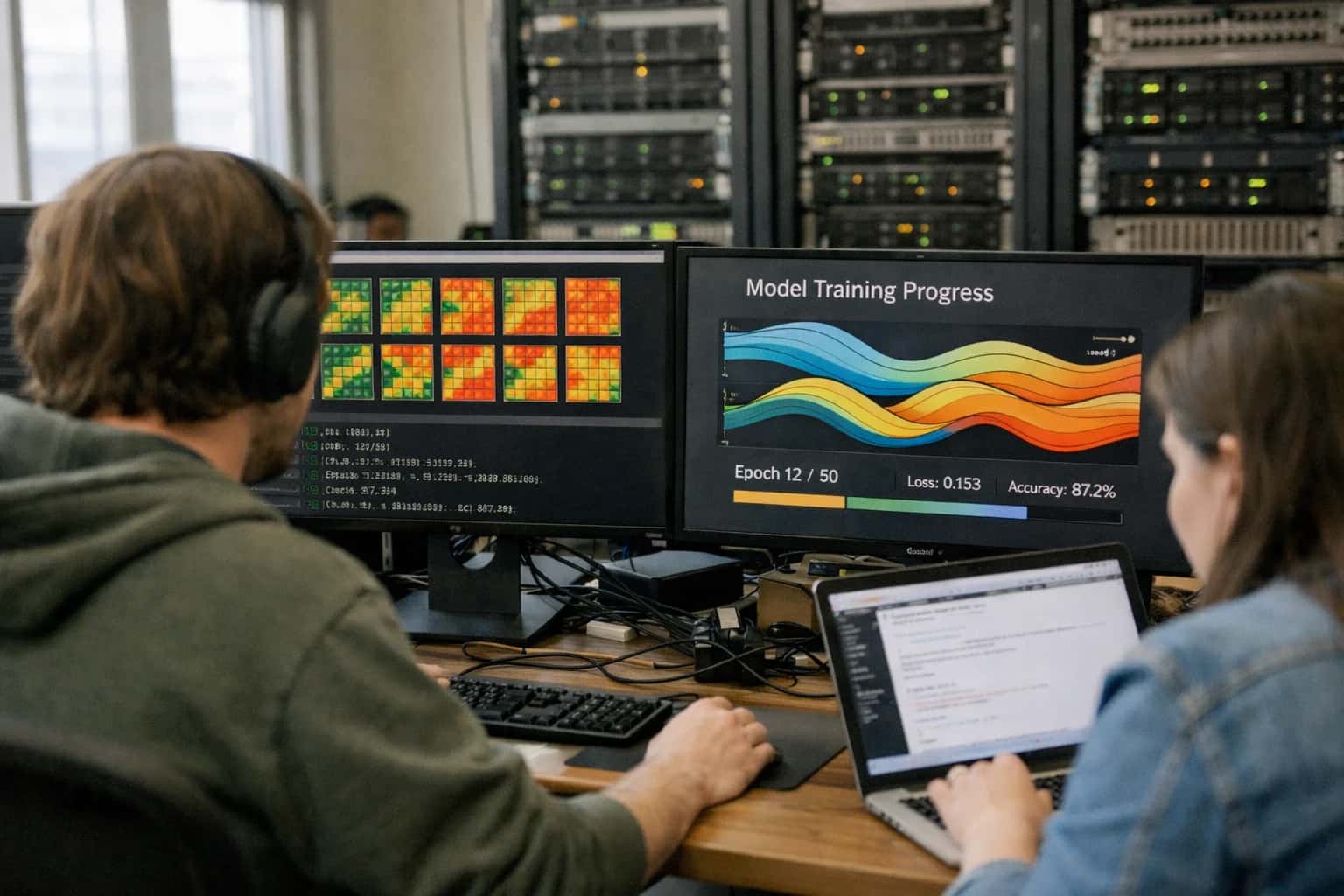
IMAGE STANDARDIZATION
What is Cross-Scanner Harmonization?
Cross-scanner harmonization is a domain adaptation technique that standardizes the appearance of medical images acquired from different scanner vendors or acquisition protocols, creating a unified data domain for robust model training and deployment.
This process is critical for transfer learning for medical imaging, as models pre-trained on data from one institution often catastrophically fail when deployed on scans from another. Harmonization enables the creation of large, heterogeneous training datasets without the confounding factor of scanner-specific artifacts, ensuring that a diagnostic model learns clinically relevant pathology rather than vendor-specific signal characteristics.
Cross-Scanner Harmonization
Key Harmonization Techniques
Standardizing medical image appearance across scanner vendors and acquisition protocols to create a unified domain for robust model training.
01
Histogram Matching
A foundational intensity normalization technique that transforms the histogram of a source image to match a reference template. How it works: The cumulative distribution function (CDF) of the source is mapped to the CDF of a target template, effectively remapping pixel intensities. Clinical relevance: Reduces gross intensity variations between GE, Siemens, and Philips scanners before feeding images into a downstream segmentation model. Limitations: Purely intensity-based; does not correct structural or textural differences introduced by reconstruction kernels.
02
Z-Score Normalization
A per-image standardization technique that rescales intensities to have zero mean and unit variance. Formula: (x - μ) / σ, where μ and σ are the mean and standard deviation of the image's voxel intensities. Why it matters: Simple, fast, and effective as a first-pass normalization step for most cross-scanner transfer learning pipelines. Caveat: Global statistics can be skewed by large pathologies or background air, so tissue-specific or masked Z-score normalization is often preferred for brain MRI or body CT.
03
CycleGAN-Based Image Translation
An unpaired image-to-image translation framework that learns to map images from a source scanner domain to a target scanner domain without requiring paired examples. Architecture: Uses a cycle-consistency loss to ensure F(G(source)) ≈ source. Application: Transforms non-contrast CTs from Scanner A to appear as if acquired on Scanner B, harmonizing texture and contrast uptake patterns. Advantage: Operates at the pixel level, directly modifying the image appearance before it enters a diagnostic model.
04
Domain-Adversarial Neural Networks (DANN)
A feature-level harmonization approach that trains a feature extractor to produce scanner-invariant representations. Mechanism: A gradient reversal layer inserted between the feature extractor and a domain classifier forces the network to learn features that are discriminative for the task but uninformative about the scanner of origin. Benefit: The downstream diagnostic model operates on a harmonized feature space, making it robust to unseen scanner protocols without modifying raw images.
05
Hounsfield Unit (HU) Calibration
A modality-specific normalization for CT that rescales raw pixel values to standardized Hounsfield Units based on scanner calibration parameters. Key thresholds: Air = -1000 HU, Water = 0 HU, Bone = +400 to +1000 HU. Implementation: Apply a fixed window/level clipping (e.g., [-1000, 400] HU for soft tissue) followed by min-max scaling. Critical step: Ensures that tissue densities are physically meaningful and consistent across vendors before any deep learning pipeline.
06
Batch Normalization Recalibration
A test-time adaptation technique that updates the running mean and variance statistics of a pre-trained model's BatchNorm layers using target domain data. Process: Pass unlabeled target images through the model in inference mode to recompute channel-wise statistics, adapting to the new scanner's intensity distribution. Advantage: Requires no backpropagation or label access; a lightweight, inference-only harmonization step. Use case: Deploying a model trained on Siemens data directly onto GE scanner outputs in a clinical setting.
Enabling Efficiency, Speed & Accuracy
Intelligent Analysis, Decision & Execution
We build AI systems for teams that need search across company data, workflow automation across tools, or AI features inside products and internal software.
Talk to Us
Search across company data
Give teams answers from docs, tickets, runbooks, and product data with sources and permissions.
Useful when people spend too long searching or get different answers from different systems.
Enterprise searchRAGPermissions
Read more
Automate internal workflows
Use AI to route work, draft outputs, trigger actions, and keep approvals and logs in place.
Useful when repetitive work moves across multiple tools and teams.
AI agentsWorkflow automationGovernance
Read more
Add AI to products and internal tools
Build assistants, guided actions, or decision support into the software your team or customers already use.
Useful when AI needs to be part of the product, not a separate tool.
AI integrationDecision supportModel routing
Read moreCROSS-SCANNER HARMONIZATION
Frequently Asked Questions
Addressing the critical challenge of domain shift caused by hardware variability, these answers explain the techniques used to standardize medical images across different scanner vendors and acquisition protocols.
Cross-scanner harmonization is a computational technique that standardizes the appearance of medical images acquired from different MRI machines or CT scanners to mitigate domain shift. It is critical because deep learning models trained on data from a single scanner vendor often fail catastrophically when deployed on images from a different manufacturer or acquisition protocol. The root cause is that neural networks exploit subtle pixel-intensity variations introduced by hardware-specific magnetic field inhomogeneities or reconstruction algorithms rather than learning the underlying pathology. Without harmonization, a model might associate a specific texture pattern with a disease simply because that texture is an artifact of a specific scanner, not a biological reality. This process creates a unified, vendor-agnostic data representation that allows a single diagnostic model to generalize robustly across a hospital network's heterogeneous fleet of imaging devices, directly enabling scalable clinical deployment and regulatory clearance.

About the author
Prasad Kumkar
CEO & MD, Inference Systems
Prasad Kumkar is the CEO & MD of Inference Systems and writes about AI systems architecture, LLM infrastructure, model serving, evaluation, and production deployment. Over 5+ years, he has worked across computer vision models, L5 autonomous vehicle systems, and LLM research, with a focus on taking complex AI ideas into real-world engineering systems.
His work and writing cover AI systems, large language models, AI agents, multimodal systems, autonomous systems, inference optimization, RAG, evaluation, and production AI engineering.
LinkedIn
Limited slotsGet a Free AI Consultation
Partnered with leading AI, data, and software stack.





How We Work
Custom AI workflows for your Business
One-fit-all AI don't work for modern businesses. At Inferensys, we aim to understand your business & custom requirements; which we use to define most efficient agentic workflows, the data, and the tools for your business.
01
Review the use case
We understand the task, the users, and where AI can actually help.
Read more02
Pick the right approach
We define what needs search, automation, or product integration.
Read more03
Build the first useful version
We implement the part that proves the value first.
Read more04
Improve from there
We add the checks and visibility needed to keep it useful.
Read moreThe first call is a practical review of your use case and the right next step.
Talk to Us