Patch-based analysis is a computational strategy where a gigapixel or high-resolution medical image is systematically divided into smaller, fixed-size sub-images called patches. Each patch is processed independently by a deep convolutional neural network to extract localized features, such as microcalcifications or spiculation patterns, that would be diluted if the entire image were downsampled to fit GPU memory constraints.

Glossary
Patch-Based Analysis
A deep learning strategy where a large mammogram is divided into smaller, overlapping sub-images for high-resolution feature extraction before global aggregation.
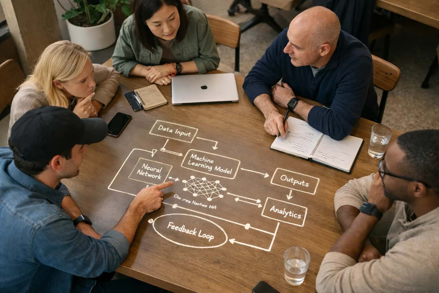
HIGH-RESOLUTION FEATURE EXTRACTION
What is Patch-Based Analysis?
A deep learning strategy where a large mammogram is divided into smaller, overlapping sub-images for high-resolution feature extraction before global aggregation.
After local feature extraction, the independent patch-level predictions are spatially aggregated using a global aggregation model to reconstruct a whole-image diagnosis. This technique is critical for whole slide image analysis and digital breast tomosynthesis, enabling models to detect subtle architectural distortions and regions of interest without sacrificing the fine-grained resolution required for clinically sensitive detection tasks.
HIGH-RESOLUTION FEATURE EXTRACTION
Key Characteristics of Patch-Based Analysis
Patch-based analysis is a foundational deep learning strategy for processing gigapixel medical images. By decomposing a full mammogram into smaller, overlapping sub-images, models can extract fine-grained textural and morphological features that would be lost in a downsampled global view.
01
Tiling and Overlap Strategy
The full-resolution mammogram is systematically divided into a grid of patches (e.g., 256x256 or 512x512 pixels). A critical parameter is the overlap stride, which ensures that lesions bisected by a tile boundary are fully captured in at least one patch. Typical overlap ranges from 10% to 50%, balancing computational load against detection sensitivity for small objects like microcalcification clusters.
02
High-Resolution Feature Preservation
Downsampling a 4000x5000 pixel mammogram to fit a standard network input destroys subtle indicators of malignancy. Patch-based analysis preserves native spatial resolution, allowing a deep convolutional neural network to learn discriminative features such as:
- Fine spiculation radiating from mass margins
- Individual microcalcification morphology and pleomorphism
- Subtle architectural distortion patterns in dense tissue
03
Global Context Aggregation
Individual patch predictions are not the final output. A global aggregation module synthesizes patch-level features into a whole-image decision. Techniques include:
- Multiple Instance Learning (MIL): Treats the image as a bag of patches, learning which patches are diagnostically relevant
- Spatial pooling: Combines feature vectors from all patches before a final classifier
- Recurrent aggregation: Sequentially processes patches to model spatial dependencies across the breast
04
Class Imbalance Handling
In a typical mammogram, over 99% of extracted patches represent normal tissue. This extreme class imbalance can cause models to predict 'normal' universally. Mitigation strategies include:
- Hard negative mining: Actively selecting patches the model finds difficult to classify correctly
- Focal loss: A loss function that down-weights easy negative examples, forcing the model to focus on rare positive patches containing lesions
- Patch-level data augmentation: Oversampling minority class patches during training
05
Computational Efficiency Trade-offs
Processing thousands of overlapping patches per image is computationally intensive. Optimizations include:
- Fully convolutional networks: Replace dense layers to process arbitrary patch sizes efficiently
- Tissue masking: Pre-segment the breast region to skip background patches entirely
- Cascaded architectures: A fast, low-sensitivity model screens all patches; only suspicious candidates pass to a slower, high-accuracy model These techniques are essential for meeting clinical worklist prioritization latency requirements.
06
Multi-Resolution Patch Pyramids
A single patch size cannot capture both fine calcifications and large masses. Multi-resolution analysis extracts patches at multiple scales:
- High-resolution patches (small field of view): Capture microcalcification morphology
- Low-resolution patches (large field of view): Capture mass margins and surrounding architectural distortion Feature vectors from each scale are concatenated or fused via attention mechanisms, providing the model with both local texture and regional context.
PATCH-BASED ANALYSIS
Frequently Asked Questions
Addressing common technical questions about the patch-based deep learning strategy used for high-resolution mammography analysis.
Patch-based analysis is a deep learning strategy where a high-resolution mammogram is systematically divided into a grid of smaller, overlapping sub-images called patches. Instead of processing the entire image at once—which would exceed GPU memory and lose fine detail—a convolutional neural network (CNN) extracts high-resolution features from each patch independently. These local features are then aggregated through a global pooling or transformer mechanism to form a whole-image diagnosis. This approach allows the model to detect microcalcifications and subtle architectural distortions that would be invisible at a downsampled resolution.
Enabling Efficiency, Speed & Accuracy
Intelligent Analysis, Decision & Execution
We build AI systems for teams that need search across company data, workflow automation across tools, or AI features inside products and internal software.
Talk to Us
Search across company data
Give teams answers from docs, tickets, runbooks, and product data with sources and permissions.
Useful when people spend too long searching or get different answers from different systems.
Enterprise searchRAGPermissions
Read more
Automate internal workflows
Use AI to route work, draft outputs, trigger actions, and keep approvals and logs in place.
Useful when repetitive work moves across multiple tools and teams.
AI agentsWorkflow automationGovernance
Read more
Add AI to products and internal tools
Build assistants, guided actions, or decision support into the software your team or customers already use.
Useful when AI needs to be part of the product, not a separate tool.
AI integrationDecision supportModel routing
Read more
About the author
Prasad Kumkar
CEO & MD, Inference Systems
Prasad Kumkar is the CEO & MD of Inference Systems and writes about AI systems architecture, LLM infrastructure, model serving, evaluation, and production deployment. Over 5+ years, he has worked across computer vision models, L5 autonomous vehicle systems, and LLM research, with a focus on taking complex AI ideas into real-world engineering systems.
His work and writing cover AI systems, large language models, AI agents, multimodal systems, autonomous systems, inference optimization, RAG, evaluation, and production AI engineering.
LinkedIn
Limited slotsGet a Free AI Consultation
Partnered with leading AI, data, and software stack.





How We Work
Custom AI workflows for your Business
One-fit-all AI don't work for modern businesses. At Inferensys, we aim to understand your business & custom requirements; which we use to define most efficient agentic workflows, the data, and the tools for your business.
01
Review the use case
We understand the task, the users, and where AI can actually help.
Read more02
Pick the right approach
We define what needs search, automation, or product integration.
Read more03
Build the first useful version
We implement the part that proves the value first.
Read more04
Improve from there
We add the checks and visibility needed to keep it useful.
Read moreThe first call is a practical review of your use case and the right next step.
Talk to Us