Spatial domain detection is an unsupervised machine learning task that identifies contiguous tissue regions with homogeneous molecular signatures. By integrating spatial transcriptomics data with histology images, algorithms such as spatial graph neural networks and spatial hidden Markov models cluster spots or cells that share similar gene expression patterns while respecting physical proximity, revealing functional anatomical structures without relying on prior manual annotations.

Glossary
Spatial Domain Detection
The unsupervised identification of tissue regions with coherent gene expression profiles and histology, often achieved through graph-based clustering or hidden Markov models.
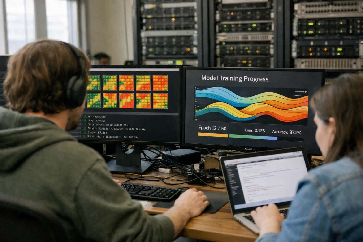
TISSUE ARCHITECTURE ANALYSIS
What is Spatial Domain Detection?
Spatial domain detection is the unsupervised computational process of partitioning a tissue section into distinct, biologically meaningful regions based on coherent gene expression profiles and histological features.
These methods construct a spatial neighborhood graph where nodes represent capture locations and edges encode adjacency, enabling the model to enforce spatial smoothness in its clustering. The output is a tissue segmentation map that directly links molecular identity to tissue architecture, serving as a critical foundation for downstream analyses like spatial differential expression, ligand-receptor co-localization, and spatial niche analysis.
UNSUPERVISED TISSUE REGION IDENTIFICATION
Key Characteristics of Spatial Domain Detection
Spatial domain detection algorithms partition tissue into functionally coherent regions by jointly modeling gene expression and spatial adjacency, revealing anatomical structures without prior histological annotation.
01
Graph-Based Clustering Foundations
The core mechanism constructs a spatial neighborhood graph where nodes represent capture locations (spots or cells) and edges connect k-nearest spatial neighbors. Clustering algorithms like Leiden or Louvain then partition this graph to maximize intra-domain expression similarity while respecting spatial contiguity. This approach enforces that domains are not just transcriptionally similar but also physically connected, preventing fragmented or biologically implausible regions.
02
Hidden Markov Random Field Modeling
A probabilistic framework that assumes each spatial location has an unobserved hidden state (the domain label) and that the observed gene expression depends on this state. Crucially, the model incorporates a Markov assumption: the domain label at one location depends only on its immediate neighbors. Methods like SpaGCN and BayesSpace use this to smooth noisy expression data, enhancing domain boundaries that align with histological structures while suppressing technical artifacts.
03
Multi-Modal Feature Integration
Advanced detectors fuse heterogeneous data streams to improve domain accuracy:
- Gene expression: Normalized count matrices from spatial transcriptomics
- Histology images: Pixel-level features extracted via pre-trained convolutional neural networks
- Spatial coordinates: Physical x,y positions encoding tissue architecture Joint latent space models learn shared representations where domains are defined by both molecular and morphological coherence, critical for identifying boundaries invisible to expression data alone.
04
Deep Graph Neural Network Approaches
Methods like STAGATE and SpaceFlow employ graph attention autoencoders that learn low-dimensional embeddings capturing both gene expression and spatial context. The network aggregates information from neighboring nodes through message-passing layers, producing spatially-aware latent representations. These embeddings are then clustered to identify domains. The deep learning advantage lies in capturing non-linear spatial dependencies and complex tissue architectures that linear methods miss.
05
Spatial Autocorrelation Metrics for Validation
Domain quality is quantitatively assessed using spatial statistics:
- Moran's I: Measures whether domain assignments exhibit significant spatial clustering versus random distribution
- Geary's C: Evaluates local spatial autocorrelation, sensitive to sharp boundaries
- Adjusted Rand Index: Compares detected domains against expert histological annotations when ground truth exists High Moran's I values confirm that domains are not merely transcriptional clusters but genuinely spatially coherent structures.
06
Resolution and Scale Sensitivity
Domain detection is fundamentally scale-dependent. The number of domains identified is controlled by clustering resolution parameters or the number of hidden states in probabilistic models. Fine resolutions reveal subtle sub-domains like cortical layers, while coarse resolutions identify major anatomical divisions. Multi-scale approaches iteratively detect domains at varying granularities, building hierarchical tissue atlases that capture both broad regions and fine-grained functional units.
SPATIAL DOMAIN DETECTION
Frequently Asked Questions
Clear answers to common questions about the computational identification of tissue regions with coherent gene expression and histological features.
Spatial domain detection is the unsupervised computational process of partitioning a tissue section into distinct, biologically meaningful regions based on coherent gene expression profiles and histological features. It works by constructing a spatial neighborhood graph where each node represents a capture spot or cell, and edges encode physical proximity. Algorithms then apply graph-based clustering (such as Leiden or Louvain community detection) or spatial hidden Markov models to group locations that share similar transcriptomic signatures while respecting spatial continuity. Unlike non-spatial clustering, these methods explicitly penalize spatially fragmented assignments, ensuring that identified domains are contiguous tissue structures like cortical layers, tumor microenvironments, or developmental zones. The output is a spatial map where each location is assigned a domain label, enabling downstream analyses such as differential expression between regions or ligand-receptor interaction mapping across domain boundaries.
Unsupervised Tissue Architecture Discovery
Computational Methods for Spatial Domain Detection
Computational methods for spatial domain detection identify tissue regions with coherent gene expression profiles and histological features. These unsupervised algorithms partition spatial transcriptomics data into anatomically meaningful compartments without prior annotation.
01
Graph-Based Clustering
Constructs a spatial neighborhood graph where nodes represent spots or cells and edges connect spatial neighbors. Algorithms like Louvain or Leiden community detection then partition this graph to identify domains with similar transcriptomic profiles.
- Leverages both gene expression similarity and physical adjacency
- Scales efficiently to datasets with hundreds of thousands of spatial locations
- Commonly used in tools like Seurat, Scanpy, and Squidpy
- Graph construction parameters (k-nearest neighbors, distance thresholds) critically influence domain granularity
Leiden
Preferred Algorithm
k-NN
Graph Construction
02
Spatial Hidden Markov Models
Probabilistic models that assume observed gene expression at each location depends on an unobserved hidden state representing a spatial domain. Transitions between states follow a Markov random field, enforcing spatial smoothness.
- Models spatial dependencies explicitly through transition probability matrices
- BayesSpace uses a t-distributed error model with spatial priors for subspot resolution enhancement
- Naturally handles uncertainty quantification for domain assignments
- Particularly effective when tissue architecture follows smooth anatomical gradients
BayesSpace
Key Implementation
Subspot
Resolution Enhancement
03
Spatial Deep Learning
Neural network architectures that learn domain representations by integrating spatial context directly into the model. Spatial graph neural networks and convolutional autoencoders capture complex, non-linear relationships between gene expression and tissue architecture.
- SpaGCN combines graph convolution with attention mechanisms to identify domains
- STAGATE uses graph attention autoencoders for spatially aware dimensionality reduction
- Can incorporate histological image features alongside transcriptomic data
- Requires careful regularization to avoid overfitting to technical noise
SpaGCN
Graph Convolution
STAGATE
Attention Autoencoder
04
Non-Negative Matrix Factorization
Decomposes the spatial expression matrix into metagenes or factors that capture co-expression signatures associated with specific tissue domains. Each spatial location is represented as a weighted combination of these factors.
- Spatial NMF variants incorporate spatial smoothness penalties on factor loadings
- Produces interpretable factors that often correspond to known cell types or tissue structures
- Giotto implements spatial-aware NMF with HMRF priors
- Enables soft domain assignments where spots can belong partially to multiple regions
Giotto
Implementation
Soft
Assignment Type
05
Gaussian Process Models
Bayesian non-parametric models that place Gaussian process priors over spatial coordinates to model smoothly varying gene expression patterns. Domains emerge from shared latent spatial functions.
- SpatialDE uses Gaussian process regression to identify spatially variable genes as a precursor to domain detection
- GPcounts models discrete count data with Gaussian process likelihoods
- Provides rigorous uncertainty quantification through posterior distributions
- Computationally intensive for large-scale datasets but highly statistically principled
SpatialDE
Key Method
Bayesian
Framework
06
Multi-Modal Integration Methods
Algorithms that jointly analyze spatial transcriptomics with histology images, immunofluorescence, or spatial proteomics to define domains. These methods learn shared latent representations across modalities.
- MUSE integrates morphology features from H&E images with gene expression
- Spatial-MultiOmics frameworks align multiple spatial assays to a common coordinate system
- Deep learning fusion layers combine image embeddings with transcriptomic features
- Improves domain boundary detection where transcriptomic signals are subtle but histological transitions are sharp
MUSE
Morphology Integration
H&E + RNA
Common Modalities
COMPARATIVE ANALYSIS
Spatial Domain Detection vs. Related Concepts
Distinguishing spatial domain detection from adjacent analytical tasks in spatial transcriptomics
| Feature | Spatial Domain Detection | Tissue Segmentation | Spatial Autocorrelation |
|---|---|---|---|
Primary objective | Identify tissue regions with coherent gene expression profiles | Partition tissue image into anatomical regions based on pixel features | Measure statistical dependence of a variable across spatial locations |
Input data type | Gene expression matrix + spatial coordinates | Histology image (H&E, IF) | Single gene expression vector + coordinates |
Core methodology | Graph-based clustering, hidden Markov models | Convolutional neural networks, pixel classification | Moran's I, Geary's C, variograms |
Output | Discrete spatial domains with transcriptomic signatures | Labeled tissue compartments (e.g., tumor, stroma, necrosis) | Scalar statistic quantifying clustering strength |
Unsupervised learning | |||
Multi-gene integration | |||
Identifies novel regions | |||
Requires spatial neighborhood graph |
Enabling Efficiency, Speed & Accuracy
Intelligent Analysis, Decision & Execution
We build AI systems for teams that need search across company data, workflow automation across tools, or AI features inside products and internal software.
Talk to Us
Search across company data
Give teams answers from docs, tickets, runbooks, and product data with sources and permissions.
Useful when people spend too long searching or get different answers from different systems.
Enterprise searchRAGPermissions
Read more
Automate internal workflows
Use AI to route work, draft outputs, trigger actions, and keep approvals and logs in place.
Useful when repetitive work moves across multiple tools and teams.
AI agentsWorkflow automationGovernance
Read more
Add AI to products and internal tools
Build assistants, guided actions, or decision support into the software your team or customers already use.
Useful when AI needs to be part of the product, not a separate tool.
AI integrationDecision supportModel routing
Read more
About the author
Prasad Kumkar
CEO & MD, Inference Systems
Prasad Kumkar is the CEO & MD of Inference Systems and writes about AI systems architecture, LLM infrastructure, model serving, evaluation, and production deployment. Over 5+ years, he has worked across computer vision models, L5 autonomous vehicle systems, and LLM research, with a focus on taking complex AI ideas into real-world engineering systems.
His work and writing cover AI systems, large language models, AI agents, multimodal systems, autonomous systems, inference optimization, RAG, evaluation, and production AI engineering.
LinkedIn
Limited slotsGet a Free AI Consultation
Partnered with leading AI, data, and software stack.





How We Work
Custom AI workflows for your Business
One-fit-all AI don't work for modern businesses. At Inferensys, we aim to understand your business & custom requirements; which we use to define most efficient agentic workflows, the data, and the tools for your business.
01
Review the use case
We understand the task, the users, and where AI can actually help.
Read more02
Pick the right approach
We define what needs search, automation, or product integration.
Read more03
Build the first useful version
We implement the part that proves the value first.
Read more04
Improve from there
We add the checks and visibility needed to keep it useful.
Read moreThe first call is a practical review of your use case and the right next step.
Talk to Us